Real-time 3D imaging for biological and medical research
Observing biological processes in real-time, within living cells, is a key step to the detailed understanding of cell behaviour and the way in which cells interact with the world around them. However, real time 3D imaging of a single cell is experimentally challenging with current techniques, such as confocal and multi-photon microscopy, which traditionally capture sequential two-dimensional (2D) images to build a three-dimensional (3D) image.
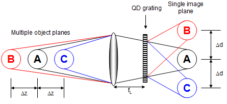
We have developing a new approach to four-dimensional (4D = 3D plus time) bio-imaging using a specially designed diffraction grating to simultaneously record information from multiple object planes onto a single image plane [1-3]. The grating behaves as a lens with a different focal length in each diffraction order, producing pseudo 3D imaging over the imaged field. The focal length of the grating, and therefore the separation of the imaged planes, is set by the design of the grating in combination with the objective magnification. This allows us to potentially match the dynamic range of the process we wish to observe to the size of the focal volume sampled by the grating (the focal volume is the image field x the distance between the two planes imaged in the ±1 diffraction orders).

A specially designed quadratically distorted grating has a different focal length in each diffraction order. Combining this with a lens provides simultaneous imaging of multiple object planes onto a single image plane. How? - Since image distance 'v' is kept constant, according to the lens equation if the focal length varies the object distance 'u' must also vary. The lens and grating are separated by distance 's' which should be set to be equal to the focal length of the lens 'fL' to provide equal magnification in the multi-plane images [1,4,5].
To see how this system is implemented on a commercial microscope system for live-cell imaging, click here. This technique is described in detail in our training document (see further reading).
References and suggestions for further reading...
We have also produced a training document designed to be both a tutorial for the complete beginner and a handy reference guide for those already familiar with the diffraction grating multiplane imaging technique. To request a PDF copy of "Introduction to Diffraction Gratings and their Application in 3D Imaging"[5] simply fill our our publication request form.
1. P.M. Blanchard, and A.H. Greenaway, Simultaneous multi-plane imaging with a distorted diffraction grating, Applied Optics, 38(32): p. 6692-9, (1999)
2. P.M. Blanchard, and A.H. Greenaway, Broadband simultaneous multi-plane imaging, Optics Communications, 183(1-4): p. 29-36, (2000).
3. P.M. Blanchard, D.J. Fisher, et al., Phase-diversity wave-front sensing with a distorted diffraction grating, Applied Optics, 39(35): p. 6649-6655, (2000).
4. S. Djidel, J.K. Gansel, H.I. Campbell, and A.H. Greenaway, High-speed, 3-dimensional, telecentric imaging, Optics Express, 14(18): p. 8269-8277, (2006).
5. H.I.C. Dalgarno, Introduction to Diffraction Gratings and their Application in 3D Imaging, training document, 2008.
back to top
Our Research
Find information on our current research projects, and our research interests both past and present...
Life Sciences Interface
- Introduction
- Real-time 3D imaging
- 3D Live-cell imaging
- Particle Tracking
- Sperm Motility
- Bioimage Gallery