3D Live-cell imaging
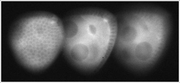
Partial view of a stage-8 Drosophila egg chamber captured using the 3D imaging system. For more details, and other live-cell images, visit our image gallery.
We are currently working with several biology and medical research groups to incorporate our diffraction grating based 3D imaging technique into live-cell imaging applications. The benefits of this new wide-field bio-imaging technique are that it is simple, robust, uses nothing but easily obtainable bulk optics components, and we have designed the system as a small module that can be adapted to fit onto many different manufacturers’ microscopes. This "bolt on" optical system basically serves as a coupling element between the microscope and the users' favourite camera.
We have designed a system which can be attached to the output camera port of our inverted Olympus IX71 microscope, but this system could be adapted to suit any microscope (inverted or upright) which delivers an image to an easily accessible camera port. The ability to obtain 'snapshot' images of several planes at once presents an advantage in dynamic imaging applications over Confocal and Multi-Photon techniques which incur a time delay by measuring the 2D slices sequentially.
The 3D imaging bolt-on simply requires that the microscope deliver an image to an optically accessible port. We have to date successfully applied this technique to provide 3D and 4D imaging with Bright Field, Phase Contrast, Dark Field, DIC and Fluorescence imaging modes (see the image gallery for examples, more are added whenever they become available!).
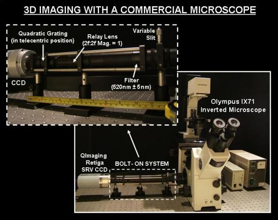
The picture above shows the current version of the bolt-on 3D imaging system installed on a commercial microscope (an Olympus IX71 inverted microscope). A lens is positioned to relay the microscope image to the CCD with 1:1 magnification. A filter is used to limit the bandwidth used (since the grating has several wavelength dependent properties, see reference [1] or request a copy of our PDF training document [2] for more on this) . We are currently developing a new version of this system that will work with broad-band illumination. A variable slit is used to limit the field of view (in one axis only) to allow three images to fit side by side on the camera and therefore make efficient use of the camera chip. The grating is placed in the 'telecentric' position (one focal length from the lens) to give equal magnification of the images [3]. Empty lens tubes have been used to bridge the gaps between each of the optical components and make the system completely light-tight. This means it can be used in normal room light avoiding the need to work in blacked out labs. This current generation bolt-on system is very robust. We have built several of these bolt-on systems and installed them on our collaborator's microscopes. Visit our image gallery to see examples of images obtained with these systems.
References and suggestions for further reading...
We have also produced a training document designed to be both a tutorial for the complete beginner and a handy reference guide for those already familiar with the diffraction grating multiplane imaging technique. To request a PDF copy of "Introduction to Diffraction Gratings and their Application in 3D Imaging"[2] simply fill our our publication request form.
1. P.M. Blanchard, and A.H. Greenaway, Broadband simultaneous multi-plane imaging, Optics Communications, 183(1-4): p. 29-36, (2000).
2. H.I.C. Dalgarno, Introduction to Diffraction Gratings and their Application in 3D Imaging, training document, 2008.
3. S. Djidel, J.K. Gansel, H.I. Campbell, and A.H. Greenaway, High-speed, 3-dimensional, telecentric imaging, Optics Express, 14(18): p. 8269-8277, (2006).
back to top
Our Research
Find information on our current research projects, and our research interests both past and present...
Life Sciences Interface
- Introduction
- Real-time 3D imaging
- 3D Live-cell imaging
- Particle Tracking
- Sperm Motility
- Bioimage Gallery